DYNAMIC VIRTUAL PATIENT
CBCT DATA INTEGRATION
Introduction
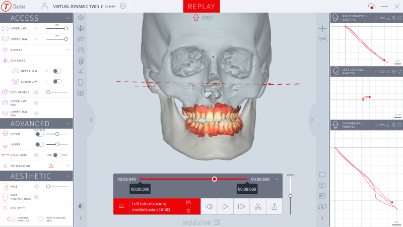
CBCT1 is widely used in dentistry, indicated in various disciplines (implantology, orthodontics, maxillofacial surgery, diagnosis of joint disorders...). TWIM™ software offers the possibility of integrating this data and building a dynamic virtual patient using patient specific motion.
How do you integrate CBCT data into the 4D workflow? Follow us through this step-by-step protocol.
Step-by-step

Step 1
CBCT ACQUISITION

Step 2
FROM DICOM TO STL

Step 3
DATA IMPORT IN TWIM
Step 1 - CBCT acquisition
The Cone Beam uses a conical X-ray beam of constant width. The principle is identical to that of conventional radiology, which practitioners use every day. The device rotates around the object under examination, projecting onto a digital sensor with each pulse. The cone beam allows the volume of the object to be obtained directly by computer calculation from the multiple 2D projections acquired during the rotation of the device. Output data is in DICOM2 format.
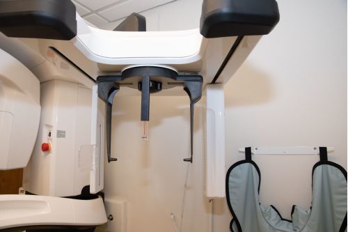

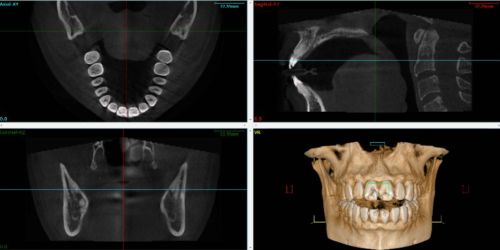
For TMJ3 analysis, wide-field scans including the entire maxilla are recommended. Moreover, file quality is directly linked to the segmentation procedure and attention must be paid to data integrity at the TMJ bone structures level
Step 2 - From DICOM to STL file
TWIM™ software and most of CAD/CAM softwares allow bone files to be imported in STL3 format, so the DICOM file must be converted into an STL one. Several open-access and fee-based softwares offer this option.
There are 3 key steps in order to import files in the correct format :
Segmentation - Conversion from DICOM to STL
Separatation of bone structures into maxilla and mandible
Matching of the 3D models from the intra-oral scanner with the bone structures.
To find out more about the conversion from DICOM to STL and how you can benefit from it, contact our team!
OPTION - Integration with ReLu®
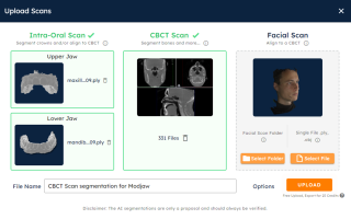
Step 1
Upload DICOM files in ReLu software
Step 2
Download results with MODJAW presets
Step 3
Upload files in TWIM software
Step 3 - Importing CBCT data in TWIM™
Segmented files are imported after creating the patient file. They are imported as maxillary and mandibular files before or after recording the patient's movements with MODJAW.
NB: before importing the CBCT files, the user must import the patient's 3D files from the intraoral camera.
- Once the file import is complete, the user is able to :
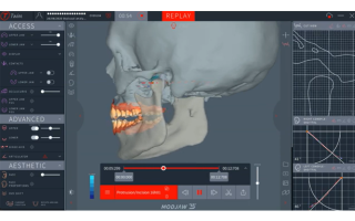
- Replay all recorded motion with CBCT data
- Modulate the appearance and transparency of the CBCT models in the "ADVANCED" module.
- Display the cut view of bone models using the “Cut view” feature
Take your analysis a step further
Track the proximity between the condylar head and the glenoid cavity during different movements.
The movements and axiographic tracings are an additional aid to the diagnosis of TMJ disorders as well as in the selection of so-called therapeutic positions.
Abréviations :
1 CBCT : Cone Beam Computed Tomography
2 DICOM : Digital Imaging and Communications in Medecine
3 ATM: Articulations temporo-mandibulaires
4 STL: Standard Triangulation Language

